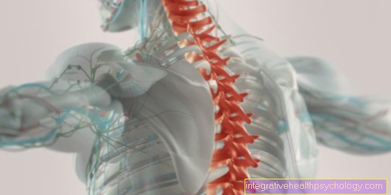
Irradiation of a heel spur (X-ray stimulus irradiation)
Synonyms
- X-ray stimulation
- Orthovolt therapy
Cause and development of a calcaneal spur
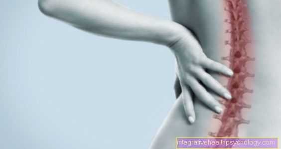
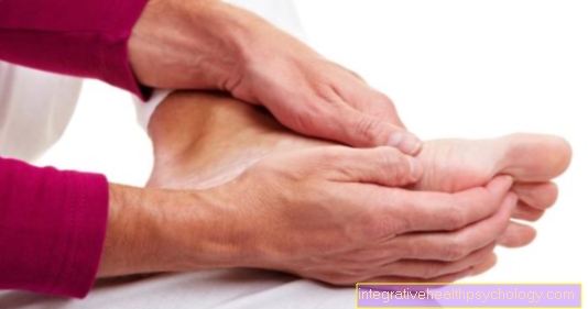
The root cause the development of heel spurs is based on increased pressure and tensile stress on the tendon attachments on the calcaneus body. This stimulus sets in motion remodeling processes in the tendon fibers, which ultimately lead to a spur-like new bone formation directed towards the foot. Of the Heel spur Due to its pressure load, it can lead to an inflammatory reaction in the surrounding tissue.
.jpg)
The triggering factors for the development of a heel spur are:
- Age
- Overweight (obesity)
- bad footwear
- Overloads (Job and sport)
- Foot malformations with extension of the longitudinal arch of the foot (often: flat arched foot, sometimes splayfoot).
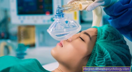
X-ray exposure
Another measure used to treat a calcaneal spur is X-ray stimulation. Often used in orthopedics after a hip prosthesis has been inserted (this is to prevent excessive bone formation), X-ray irradiation can also be used to treat a heel spur.
You can find a lot more information under our topic: X-ray stimulation
Appointment with an expert in calcaneal spur?
_2.jpg)
I would be happy to advise you!
Who am I?
My name is dr. Nicolas Gumpert. I am a specialist in orthopedics and the founder of .
Various television programs and print media report regularly about my work. On HR television you can see me every 6 weeks live on "Hallo Hessen".
But now enough is indicated ;-)
Athletes (joggers) are particularly often affected by the disease of the heel spur. In many cases, the cause of the inflammation of the heel spur cannot be identified at first. Therefore, the treatment requires a lot of experience. I focus on the heel spur.
The aim of every treatment is treatment without surgery with a complete recovery of performance.
Which therapy achieves the best results in the long term can only be determined after looking at all of the information (Examination, X-ray, ultrasound, MRI, etc.) be assessed.
You can find me in:
- Lumedis - your orthopedic surgeon
Kaiserstrasse 14
60311 Frankfurt am Main
Directly to the online appointment arrangement
Unfortunately, it is currently only possible to make an appointment with private health insurers. I hope for your understanding!
Further information about myself can be found at Dr. Nicolas Gumpert
X-rays is radioactive Radiation with accelerated particles that, depending on their strength, can penetrate tissue. Above all, less dense tissue, such as Skin and fatty tissue can be penetrated unhindered. X-rays are mostly reflected or absorbed on denser tissues. Such structures are then shown as a light area in the X-ray image. X-rays per se is cell damaging. The more intense the X-ray radiation is selected, the greater the damage the radiation can cause to the irradiated tissue. In the case of a heel spur treatment, the X-ray radiation is selected so that the surrounding tissue can be penetrated without any problems, but when it hits the heel spur tissue it carries enough energy that the bony tissue and cells irreversible be harmed. Ultimately, this leads to the fact that the bony tissue of the heel spur is increasingly broken down and becomes smaller. Sometimes repeated applications have to be carried out here as well until the desired effect can be achieved.
It is important to note that the energy of the X-ray radiation is selected so that the amount is not too damaging. Furthermore, with X-ray irradiation, unlike shock wave irradiation, greater attention must be paid to the fact that the radiation is directed directly onto the area to be irradiated, in this case the heel spur. If the area is set too generously, the surrounding, unaffected tissue, such as annoy and Blood vessels can be irreversibly damaged by the X-rays. The possible side effects from this would be Bleeding, Sensory disturbances and if necessary Nerve pain.
At times it can too irritations the skin in the area of the irradiated area. This is because the X-ray radiation must first penetrate a corresponding area of skin before it can reach the bone. This can lead to the described irritation of the skin and corresponding irritations. To minimize the side effects, you can post X-ray exposure cooling gels applied to the skin. The skin usually regenerates within a few days. After several weeks there is usually nothing left to see of the radiation.
Neither with shock wave therapy nor with X-ray irradiation, it is not the case that the bony structures have suddenly all disappeared, it is usually the case that one slow downsizing of the heel spur indicates a good response to therapy.
Irradiation process
At the first appointment the patient receives a detailed consultation, in which the attending physician is once again all Diagnostics (such as x-rays). In addition, information is provided about side effects and risks and questions from the patient are answered. The irradiation of the heel spur is done by computer programs individually to the patient adjusted so that only the heel spur region is irradiated.
Of the next appointment will the actual irradiation which usually only takes a few seconds to minutes. The irradiation is then approximate two times a week, depending on the radiation dose used for three to eight weeks carried out. After the radiation cycle, follow-up appointments are made to discuss the success of the therapy and how to proceed.
Duration of treatment
The irradiation of the heel spur lasts usually five weeks. During this time, the heel is usually exposed to low-dose radiation two times a week irradiated. The majority of patients are free of pain and symptoms after this five-week treatment. In some cases, the irradiation on heel spurs shows an effect after three weeks. However, the pain may not have gone away after the first five weeks. Then the therapy time extended to up to eight weeks. If after this treatment period no complete freedom from pain is achieved, the cycle can be repeated after two to three months.
When does the pain get better?
The freedom from pain after irradiation of the heel spur varies from patient to patient. So it can be that the pain gets better after the first or second radiation. After a cycle of three to five weeks of radiation, most patients are relieved of pain. The pain can, however, also occur during radiation stronger in the short term become. However, this should not cause concern as it is a known side effect. Pain relief of the heel spur can rarely be achieved after the first irradiation cycle, so that a further treatment sequence can be followed after a few months.
Prognosis for radiation use
The prognosis for radiation therapy for heel spurs is generally very good. About 80-90% of patients are pain-free after the first course of treatment. At about 60% the patient is the Freedom from pain after irradiation of the calcaneal spur permanent. If the irradiation in the first cycle did not have an adequate effect, a further treatment cycle must be discussed and, if necessary, followed by a break of a few weeks or months.
Since the irradiation does not cause any changes in bones or joints, but only serves to relieve pain, in rare cases pain can occur again due to recurrent inflammation of the heel spur.
Radiation Therapy Risks
Since the irradiation is a therapy with ionizing radiation, can isolated side effects occur during treatment. However, when the heel spur is irradiated, the radiation exposure is very low (about 6 gray) and only limited to a small region of the body. Hence are Side effects rare. In very rare cases and with previously damaged skin, skin irritation, rashes or reddening can occur at the site of irradiation. In addition, a temporary increase in pain is possible during the radiation.
In general, the sensitive genital organs are particularly at risk in the case of radiation, as this can change the genetic make-up. Therefore, one should weigh up whether radiation is really necessary for men or women who still want to have children. However, since radiation can nowadays be aimed very locally and because of the low doses, negative effects on the genital organs are very rare.
Before irradiating the heel spur in women, pregnancy should be excluded, as the therapy can lead to an abortion or malformations in the unborn child.
In principle, the risk of causing a tumor through irradiation of the heel spur cannot be ruled out, although this is very unlikely at the low dose.
Alternative treatment - shock wave therapy
Another non-surgical measure to treat a heel spur is shock wave therapy. Shock wave therapy is already known from the treatment of kidney stones. The mechanism is that targeted shock waves are directed onto an area of tissue. This passes the waves on to neighboring tissue, which is increasingly set in motion. As soon as the bony substance of the heel spur is reached, the molecules of the bone are also set in motion.
Initially, there is no change in the structure. The longer this type of irradiation is carried out or the more frequently the treatment is repeated, the more unstable the bony structures become. When used successfully, the heel spur becomes increasingly smaller, which increasingly degrades inwards from the edge. What causes kidney stones to disintegrate, so leads to crumbling in a heel spur.
If used correctly, the treatment is relatively painless and, in contrast to shock wave therapy for kidney stones, is described by patients as moderately uncomfortable.
The painfulness depends crucially on the practitioner and the shock wave device. The more efficient, focused shock waves are significantly less painful than the radial shock wave therapy.
Several sessions are usually necessary before the tissue reacts to the ultrasonic waves.
In the meantime, the statutory insurance pays the treatment under certain conditions. Private health insurances take over the treatment completely. If one decides for a treatment, the patient usually has to reckon with several hundred euros for several sessions.
Even after successful treatment with ultrasound waves, the formation of new heel spurs cannot be ruled out. The reason for this is that the usually strong incorrect loads that have led to the formation of the heel spur continue to be carried out and chronic overstressing of the foot is not reduced. That is why it is very important that after a treatment appropriate bad posture is corrected and excessive strain is greatly reduced. In any case, intensive physiotherapy that works out precisely these weak points should be carried out. A post-treatment period of several weeks should be expected in this context.
In contrast to shock wave therapy, the physiotherapeutic follow-up treatments are always covered by statutory health insurances. Physiotherapists usually prescribe 10-12 sessions, which must be followed consistently. Ultrasonic shock wave therapy is practically without risk and side effects. In some cases, however, tissue irritation of the foot can occur, especially if the waves are too strong.
Appointment with ?
_3.jpg)
I would be happy to advise you!
Who am I?
My name is dr. Nicolas Gumpert. I am a specialist in orthopedics and the founder of .
Various television programs and print media report regularly about my work. On HR television you can see me every 6 weeks live on "Hallo Hessen".
But now enough is indicated ;-)
Athletes (joggers, soccer players, etc.) are particularly often affected by diseases of the foot. In some cases, the cause of the foot discomfort cannot be identified at first.
Therefore, the treatment of the foot (e.g. Achilles tendonitis, heel spurs, etc.) requires a lot of experience.
I focus on a wide variety of foot diseases.
The aim of every treatment is treatment without surgery with a complete recovery of performance.
Which therapy achieves the best results in the long term can only be determined after looking at all of the information (Examination, X-ray, ultrasound, MRI, etc.) be assessed.
You can find me in:
- Lumedis - your orthopedic surgeon
Kaiserstrasse 14
60311 Frankfurt am Main
Directly to the online appointment arrangement
Unfortunately, it is currently only possible to make an appointment with private health insurers. I hope for your understanding!
Further information about myself can be found at Dr. Nicolas Gumpert
forecast
The prognosis for a successful Heel spur treatment is good. Almost always (>90%) a clear symptom relief or freedom from symptoms is achieved. The success of the therapy depends, among other things, on the possibility of physical rest during the treatment period. Since this is seldom possible, phases of suffering lasting several months to several years often arise.
The clinical picture of Heel spur also tends to Relapse (renewed complaints). Even if a conservative or operative therapy Successful recurrence symptoms can occur at any time.
Read more on the topic Voltaren Dispers at our partner.
Figure heel spur
_4.jpg)
Heel spur - calcaneus spur
(bony outgrowth)
- Lower (plantar) heel spur
- Upper (dorsal) heel spur
- Calcaneal tuberosity -
Calcaneal tuberosity - Heel bone - Calcaneus
- Achilles tendon -
Tendo calcaneus - External calf muscle -
M. gastrocnemius,
Caput laterale - Shin - Tibia
- Fibula - Fibula
- Ankle bone - Talus
- Scaphoid bone - Navicular bone
- External sphenoid bone -
Os cuneiform laterale - Cuboid bone - Os cuboideum
- Sole tendon plate -
Plantar aponeurosis
A - picture of heel with a lower one
and upper calcaneal spur
B - Left foot from the outside
You can find an overview of all Dr-Gumpert images at: medical illustrations


.jpg)